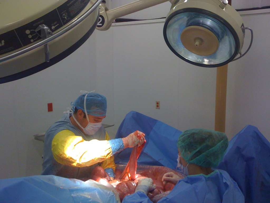
CALEC surgery represents a groundbreaking advancement in the field of eye damage treatment, specifically targeting blinding corneal injuries. This innovative procedure, initiated at Mass Eye and Ear, utilizes stem cell therapy to restore the cornea’s surface by cultivating autologous limbal epithelial cells. During a clinical trial, the effectiveness of this surgical method was demonstrated, revealing that over 90 percent of participants experienced significant improvements in their corneal health. As we delve deeper into CALEC surgery, we uncover a ray of hope for those suffering from corneal damage previously deemed untreatable, thanks to the pioneering efforts of researchers like Ula Jurkunas. Moreover, the implications of CALEC surgery extend beyond mere restoration; they evoke a new era of potential treatments for various eye conditions, igniting optimism within the ophthalmology community and among patients alike.
Cultivated autologous limbal epithelial cell (CALEC) therapy emerges as a revolutionary approach in ocular medicine, specifically aimed at addressing severe corneal injuries. This cutting-edge surgical technique involves extracting healthy stem cells from a donor eye and meticulously regenerating them into tissue grafts that can be transplanted into a damaged cornea. With roots at esteemed institutions like Mass Eye and Ear, CALEC provides renewed hope for individuals enduring vision loss due to conditions such as chemical burns or infections. Enhancements in corneal repair techniques like CALEC underscore the importance of limbal epithelial cells in maintaining ocular health and restoring sight. As the medical community further explores this procedure, the potential for improved outcomes in eye damage treatment is promising, paving the way for future advancements in regenerative eye therapies.
Understanding CALEC Surgery and Its Implications for Eye Healing
CALEC surgery, or Cultivated Autologous Limbal Epithelial Cell surgery, offers groundbreaking hope for patients suffering from severe corneal damage. Conducted by Ula Jurkunas and her team at Mass Eye and Ear, this innovative procedure involves extracting healthy limbal epithelial cells from a patient’s unaffected eye, cultivating them into a graft, and transplanting them to restore the cornea’s damaged surface. With a striking efficacy rate of over 90% in initial trials, CALEC not only improves vision but also alleviates related pain and discomfort, providing a new avenue for damage previously deemed untreatable.
The success of CALEC surgery hinges on the significant role that limbal epithelial cells play in maintaining corneal integrity. These cells are crucial for the regeneration of the cornea’s surface, which can be compromised due to injuries like chemical burns or infections. This procedure represents a shift in the treatment paradigm, offering patients a viable alternative to traditional methods that may not yield successful outcomes. As research progresses, it holds the potential for wider applications, particularly in those who may have sustained damage to both eyes.
The Role of Stem Cell Therapy in Corneal Repair
Stem cell therapy has emerged as a revolutionary approach in the field of ocular regeneration, particularly for corneal repair. The recent clinical trials at Mass Eye and Ear focused on using cultivated autologous limbal epithelial cells (CALEC) demonstrate how taking stem cells from a patient’s healthy eye can effectively combat severe corneal injuries. This innovative type of treatment not only restores the corneal surface but also re-empowers patients who had lost hope for restoration due to the degree of their injuries.
By utilizing a patient’s own stem cells, the risks often associated with allogeneic transplants are mitigated. This ensures a higher compatibility and lower rejection rates, thus enhancing the healing process. With the promising results seen in the CALEC studies, researchers are encouraged to explore expanding these biological therapies further, potentially developing techniques that could allow bilateral application, making groundbreaking strides in corneal repair and redefining eye damage treatment.
Regeneration of Limbal Epithelial Cells: A Game Changer
The regeneration of limbal epithelial cells stands at the forefront of eye care innovations, presenting a game-changing solution for individuals suffering from blinding corneal injuries. The approach taken in the CALEC surgery not only addresses the restoration of the cornea but also highlights the significant role these cells play in overall ocular health. As it stands, patients who experience severe damage to their cornea often face limited options, and the depletion of limbal cells drastically reduces the chances of regeneration.
Innovative therapies, such as those developed at Mass Eye and Ear, aim to replenish these crucial cells by enabling patients to harness their own biological materials for healing. This breakthrough has opened new avenues for research, paving the way for larger trials and potentially fulfilling the unmet needs of a substantial patient population facing vision loss due to corneal failure. The implications could be profound, not just improving individual patient outcomes but also advancing the field of eye care through enhanced therapeutic options.
Clinical Trial Success: A Look into CALEC Outcomes
The clinical trials surrounding the CALEC (Cultivated Autologous Limbal Epithelial Cells) procedure have produced remarkable outcomes, showcasing its potential as an effective treatment for corneal injuries. Out of 14 participants in the initial study, the results showed a success rate of over 90% in restoring the corneal surface after treatment, with follow-up evaluations indicating continued improvements in visual acuity. The trial’s design and its coordination with cell manufacturing processes reflect a significant advancement in the medical community’s approach to stem cell therapy.
These outcomes underscore the importance of rigorous clinical trials in establishing the safety and effectiveness of new treatments. Throughout the trials, no severe adverse events were noted, further validating the procedure’s safety profile. As this research continues to evolve, it presents a hopeful narrative for future treatments and reflects the strides being made in regenerative medicine for eye damage treatment.
Future Directions for CALEC and Ocular Regeneration Research
As the research surrounding CALEC surgery progresses, there is an optimistic outlook for the future of ocular regeneration therapies. The current trials have provided invaluable data, not only in assessing the effectiveness of stem cell strategies but also in paving the way for additional studies aimed at broadening the scope of treatment for corneal damage. Future cohorts will involve larger patient populations and extended follow-ups, essential for gaining enough evidence to support regulatory approval.
The aspiration to establish an allogeneic manufacturing process using donor limbal stem cells will also be a significant milestone, as it could democratize access to this revolutionary treatment and make it available to a wider patient demographic. By continuing to push the boundaries of stem cell research and clinical applications, researchers are positioning themselves to transform the landscape of eye care and offer hope to countless individuals suffering from corneal injuries.
The Importance of Limbal Cell Preservation in Eye Health
Limbal cell preservation is central to maintaining eye health, as these cells play an essential role in the regeneration of the cornea’s surface. When limbal epithelial cells are lost due to injury or disease, the risk of complications increases significantly, leading to a myriad of visual impairments. Understanding this, the CALEC approach highlights the crucial need for effective strategies to preserve and regenerate these cells, ensuring the cornea remains functional.
Protecting and utilizing these valuable cells can revolutionize the management of corneal injuries. As more attention is drawn towards the significance of limbal stem cells in ocular health, interventions like the CALEC surgery could lead to revolutionary changes in how clinicians approach the restoration of vision for patients battling severe eye damage and chronic conditions.
The Safety Profile of CALEC Surgery
One of the most promising attributes of CALEC surgery is its robust safety profile, as evidenced by the clinical trial data gathered from participants. The absence of any severe adverse events reinforces the notion that this stem cell therapy can be deployed with relatively low risk. The minor complications reported, such as a localized bacterial infection, are manageable and often resolve without extensive intervention, making the procedure viable for more patients suffering from corneal injuries.
This high level of safety is essential not only for patient reassurance but also for the broader acceptance and potential regulatory approval of the therapy. As researchers work towards optimizing and refining CALEC protocols, the emphasis on ensuring patient safety will likely remain a top priority, reinforcing trust in innovative ocular treatments and paving the way for more widespread clinical adoption.
Collaboration in Vision Research: A Collective Effort
The success of CALEC surgery at Mass Eye and Ear illustrates the power of collaboration in advancing vision research. The concerted efforts of various institutions, including Dana-Farber Cancer Institute and Boston Children’s Hospital, exemplify how interdisciplinary cooperation can lead to meaningful innovations in treatment. By combining expertise across scientific, clinical, and engineering fields, teams are able to develop and implement techniques that have far-reaching implications for patients with corneal damage.”},{
Frequently Asked Questions
What is CALEC surgery and how does it relate to eye damage treatment?
CALEC surgery, or cultivated autologous limbal epithelial cell surgery, is a groundbreaking procedure developed at Mass Eye and Ear for treating severe eye damage, particularly blinding corneal injuries. It involves taking stem cells from a healthy eye, expanding them into grafts, and transplanting them into the damaged eye to restore its corneal surface. This innovative approach offers new hope for patients with conditions previously deemed untreatable.
How does stem cell therapy improve corneal repair in CALEC surgery?
In CALEC surgery, stem cell therapy plays a vital role in corneal repair. By harvesting limbal epithelial cells from a healthy eye and using them to create a cellular graft, the procedure effectively restores the damaged cornea. Clinical trials have shown that this stem cell treatment can achieve over 90% effectiveness in repairing the corneal surface, significantly enhancing the patient’s vision and quality of life.
What are limbal epithelial cells and their importance in CALEC surgery?
Limbal epithelial cells are crucial components in the health of the cornea, located at the limbus, which is the edge of the cornea. They ensure the smooth surface of the eye and play a key role in healing and regeneration. In CALEC surgery, these cells are harvested from a healthy eye to create a graft for transplantation into a damaged eye, making the treatment highly effective for restoring vision in patients with corneal damage.
Is CALEC surgery currently available for patients with corneal damage?
As of now, CALEC surgery remains an experimental procedure and is not widely available at Mass Eye and Ear or other hospitals in the U.S. Further studies and trials are necessary before it can be submitted for federal approval. The research team aims to conduct more extensive trials to eventually offer this innovative treatment to patients nationwide.
What were the key findings from the clinical trials of CALEC surgery at Mass Eye and Ear?
The clinical trials conducted at Mass Eye and Ear showed promising results for CALEC surgery, with a 90% overall success rate in restoring the corneal surface among participants. At follow-ups, complete restoration was observed in 50% of patients at three months, increasing to 79% and 77% at 12 and 18 months, respectively. The procedure demonstrated a high safety profile, with minimal adverse effects reported.
What makes CALEC surgery a new hope for patients suffering from eye injuries?
CALEC surgery offers a revolutionary treatment option for patients with corneal damage caused by trauma, infections, or chemical burns. Traditionally, such injuries were considered untreatable, leaving patients with persistent pain and vision loss. With over 90% efficacy in restoring corneal surfaces, CALEC surgery represents a significant advancement in eye damage treatment, providing new hope for recovery and improved quality of life.
How is the CALEC procedure performed and what is the recovery process?
The CALEC procedure consists of several steps: a biopsy is taken from a healthy eye to extract limbal epithelial cells, which are then cultured to form a graft. After a two to three-week growth period, the graft is surgically transplanted into the damaged eye. Recovery processes vary per patient, but the surgery aims to restore corneal surfaces to alleviate pain and improve vision over time, with regular follow-up assessments for progress.
What future advancements are planned for CALEC surgery?
Researchers aspire to develop an allogeneic manufacturing process using limbal stem cells from donors, which would expand CALEC surgery’s applicability to patients with damage in both eyes. Future studies aim to involve larger patient groups and explore randomized controls to solidify the treatment’s efficacy and move closer to FDA approval, ultimately making this life-changing therapy accessible to more patients.
Who leads the research and clinical trials for CALEC surgery at Mass Eye and Ear?
The research and clinical trials for CALEC surgery are led by Dr. Ula Jurkunas, an associate director of the Cornea Service at Mass Eye and Ear, alongside Dr. Reza Dana. Together, they have pioneered this innovative stem cell treatment for corneal repair, ensuring ongoing advancements in the field of eye damage treatment.
| Key Points |
|---|
| CALEC surgery, pioneered by Ula Jurkunas at Mass Eye and Ear, offers a groundbreaking solution for patients with severe corneal damage previously deemed untreatable. |
| The treatment involves harvesting stem cells from a healthy eye, expanding them into a graft, and transplanting this graft into the damaged eye. |
| In a clinical trial involving 14 patients, CALEC demonstrated over a 90% success rate in restoring corneal surfaces. |
| Follow-up results showed a 50% improvement at three months, increasing to 92% by 18 months. |
| The procedure was safe, with no severe adverse events reported, although some minor complications occurred. |
| Next steps include larger trials to broaden access and potentially treat patients with damage to both eyes. |
Summary
CALEC surgery represents a significant advancement in the treatment of corneal damage, restoring hope for patients with previously untreatable conditions. This innovative approach, utilizing stem cell therapy, has shown high success rates in clinical trials and marks a pivotal moment in ophthalmology. As research continues, further studies aim to solidify CALEC’s efficacy and expand its application, potentially offering a viable solution for individuals suffering from debilitating eye injuries.
